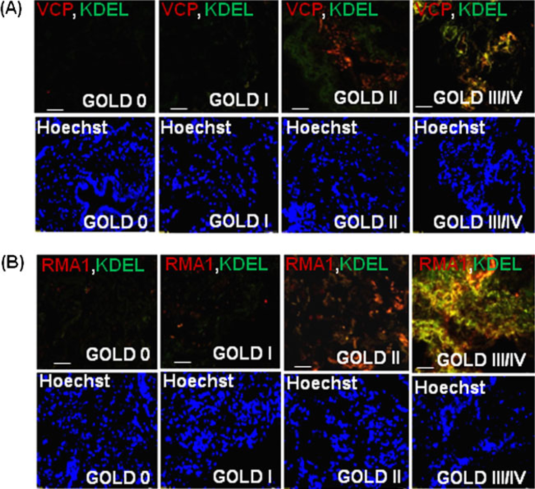
Fig. 5.
Valosin-containing protein (VCP) interacts with gp78 and Rma1 on the ER membrane. The paraffin-embedded longitudinal human lung sections of control (GOLD 0) and COPD lung tissues at GOLD I–IV levels of emphysema (n=4–5, each group), were immunostained with primary mouse monoclonal antibodies for VCP (red, a) or Rma1 (red, b). These sections were co-immunostained with rabbit polyclonal KDEL (green, ER membrane marker). a The data show the co-localization of VCP and KDEL in severe (GOLD III/IV) emphysema lung tissues compared with the mild or moderate (GOLD I/II) emphysema and GOLD 0 lung tissues. b The co-localization of Rma1 and KDEL is seen in moderate and severe (GOLD II/III/IV) emphysema lung tissues compared with the mild (GOLD I) emphysema and control (GOLD 0) lung tissues. The data not only verify the increased VCP-Rma1 expression but also demonstrate its localization in or around ER membrane with increasing severity of emphysema in COPD. The nuclear (Hoechst) staining is shown in the bottom panels. Scale=50 µm

