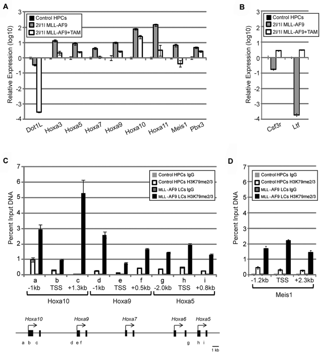
Figure 6.
DOT1L directly regulates expression of Hoxa and Meis1 genes in MLL-AF9–transformed cells. (A) RT-qPCR analysis shows up-regulation of Hoxa cluster and their cofactors Meis1 and Pbx3, normalized to Gapdh, in MLL-AF9–transformed cells, which become down-regulated on DOT1L deletion. (B) RT-qPCR analysis shows down-regulation of myeloid differentiation markers, normalized to Gapdh, in MLL-AF9–transformed cells, which become up-regulated on DOT1L deletion. (C) ChIP analysis demonstrates that H3K79me2/3 is specifically enriched at Hoxa loci in MLL-AF9–transformed cells compared with control HPCs. IgG was used for control ChIP and amplicon positions are indicated (TSS indicates transcription start site). (D) ChIP analysis demonstrates that H3K79me2/3 is enriched in the Meis1 gene in MLL-AF9 LCs compared with that in the control HPCs. IgG was used for control ChIP and amplicon positions are indicated (TSS indicates transcription start site).

