
Figure 3.
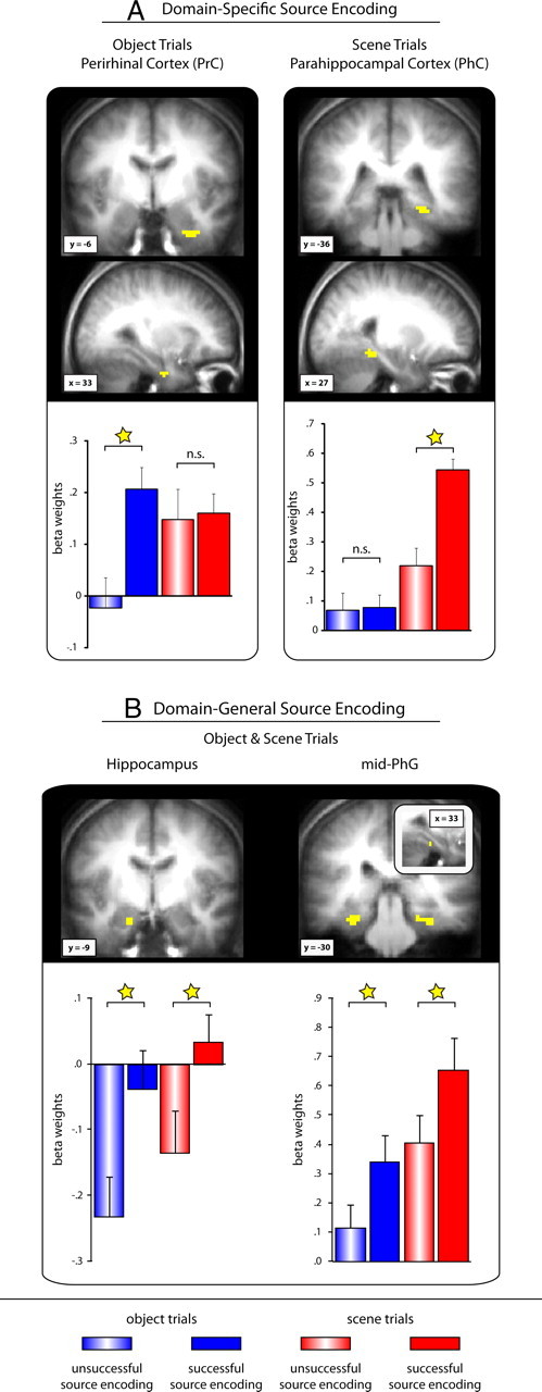
Domain-specific (A) and domain-general (B) source encoding effects in the medial temporal lobe revealed via contrast/masking procedures within the standard GLM approach. Clusters are displayed on the mean anatomical scan across subjects. The bar graphs represent mean (+SEM) β-weights across subjects (averaged across voxels within a cluster) for successful (filled bars) and unsuccessful (striped bars) object (blue) and scene (red) source encoding trials. ☆p < 0.05; n.s., not significant.