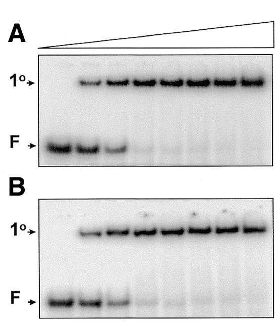
Figure 1.

Equilibrium binding. Mixtures in SfiI binding buffer contained 10 nM 32P-labelled duplex, either ATATA (A) or AAAAA (B), 1 nM pAT153 and SfiI endonuclease at one of the following concentrations (increasing from left to right across the gels, as indicated by the wedge): 0, 2.5, 5, 10, 20, 40, 80 and 160 nM. After 30 min at room temperature, the mixtures were subjected to electrophoresis through polyacrylamide. The electrophoretic mobilities of the free DNA and the primary complexes are marked on the left of the gels as F and 1°, respectively.
