FIGURE 5:
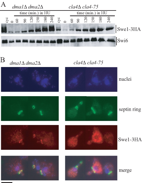
Different patterns of Swe1 stabilization in HU-treated dma1Δ dma2Δ and cla4-75 cells. Exponentially growing (cyc) cell cultures of dma1Δ dma2Δ SWE1-3HA (yRF661) and cla4-75 SWE1-3HA (yRF690) strains were arrested in G1 by α-factor at 25°C (time 0) and released from G1 arrest at 37°C in YEPD medium containing 150 mM HU. Samples were taken at the indicated times after release for determining Swe1 levels (A) as in Figure 2C and in situ immunofluorescence analysis (B) of nuclei, septin ring deposition, and Swe1-3HA localization as in Figure 4. Representative micrographs were taken at t = 180 min. Bar, 5 μm.

