
FIGURE 7:
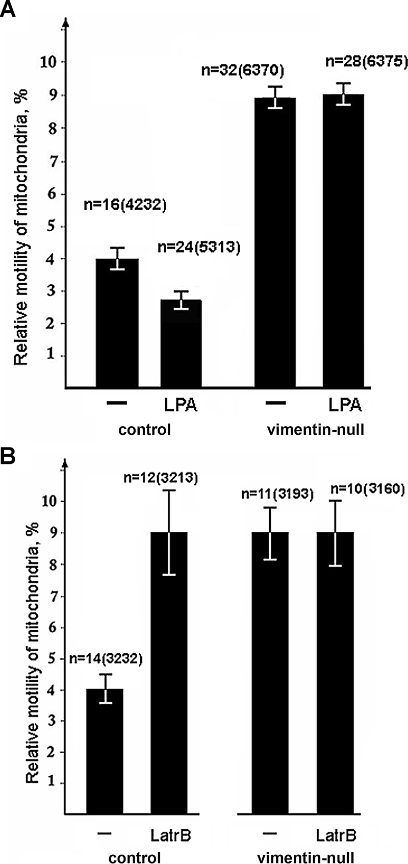
VimIFs mediate the interaction of mitochondria with F-actin. Cells were transfected with plasmid pEGFP-Mito using Maxifectin. The movements of fluorescently labeled mitochondria in normal and vimentin-null cells were recorded using time-lapse video microscopy before and after incubation with 5 μM LPA for 5 min (A) or before and after incubation with 0.2 μM Lat B for 20 min (B). Values are mean percentage of movements exceeding 0.2 μm/s ± SEM; n = number of cells, and, in brackets, number of mitochondrial movements.