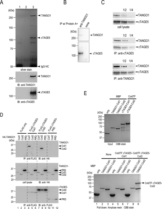
FIGURE 2:
cTAGE5 binds to TANGO1 at the ER exit sites. (A) Protein A beads conjugated with (lanes 1 and 3) or without (lane 2) anti–cTAGE5 CC1 antibody were incubated with (lanes 2 and 3) or without (lane 1) HeLa cell lysates. The beads were washed, and proteins retained to the beads were analyzed by SDS–PAGE, followed by silver staining or Western blotting with anti–cTAGE5 CC1 and TANGO1 antibodies. (B) Protein A beads were either untreated or conjugated with anti-TANGO1 antibody and then incubated with HeLa cell lysates. The beads were washed, and proteins retained to the beads were analyzed by SDS–PAGE, followed by Western blotting with anti–cTAGE5 CC1 and TANGO1 antibodies. (C) HeLa cell lysates were immunoprecipitated with anti–cTAGE5 CC1 antibody or anti-TANGO1 antibody, and immunoprecipitants and cell lysates were sequentially diluted and analyzed by SDS–PAGE and blotted with anti–cTAGE5 CC1 and anti-TANGO1 antibodies. (D) 293T cells were transfected with FLAG-tagged cTAGE5-Coil1 (amino acids 61–300), Coil2 (amino acids 301–650), or PRD (amino acids 651–804) with HA-tagged TANGO1-Coil1 (amino acids 1211–1440), Coil2 (amino acids 1441–1650), or PRD (amino acids 1651–1907). Cell lysates were immunoprecipitated with anti-FLAG antibody and eluted with FLAG peptide. Eluates and cell lysates were analyzed by SDS–PAGE, followed by Western blotting with anti-FLAG or anti-HA antibodies. (E) MBP, MBP-tagged TANGO1-Coil1, and TANGO1-Coil2 were expressed in E. coli and purified with Amylose resin. ColdTF-tagged cTAGE5-Coil1 and cTAGE5-Coil2 were expressed in E. coli and purified with Ni Sepharose. Purified proteins were analyzed by SDS–PAGE, followed by Coomassie brilliant blue (CBB) stain (top); MBP, MBP-tagged TANGO1-Coil1, and TANGO1-Coil2 were immobilized to amylose resin and untreated or incubated with ColdTF cTAGE5 Coil1 or ColdTF cTAGE5 Coil2. Resins were washed and eluted with maltose. Eluted proteins were subjected to SDS–PAGE followed by CBB stain (bottom).

