Fig. 2.
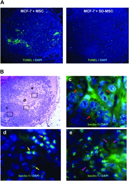
Autophagic SD-MSCs prevent apoptosis in tumor (A) MCF-7 cells were suspended in Matrigel and either injected alone or co-injected with MSCs or SD-MSCs in the mammary fat pad of SCID beige mice. After 2 weeks, tumor sections were stained for apoptotic cells using a TUNEL assay (magnification ×40). (B) Autophagy beclin-1 marker is gradually relocalized to the nucleus in the inner MCF-7 tumor. MCF-7 and SD-MSCs were co-injected in mammary fat pad of SCID beige mice. After 2 weeks, tumors were collected, sectioned and stained. Top left: hematoxylin–eosin staining of a representative section from a fully developed tumor. Top right (background set high): immunohistochemistry of beclin-1 revealed a diffuse staining in growing parts of the tumor (red arrows). Bottom left: at the frontier of the necrotic part of the tumor, beclin-1 staining is perinuclear in some cells (yellow arrows). Bottom right: in the necrotic part, most of the cells showed a relocation of beclin-1 around the nucleus (purple arrows), in addition to expressing condensed nuclei compared with the other part of the tumor. Magnification: top left panel: ×40; c, d and e: ×1000.

