
Figure 1.
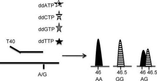
Schematic representation of the SNaP shot technique. The primers have a determined length, are specific for the genomic region where the mutant is, and end at a nucleotide preceding the mutation. Subsequently, one fluorochrome-labeled dideoxynucleotide is added. Using capillary electrophoresis, products are separated according to size. Depending on the nucleotide build up after primer extension, either one or two of the fluorochromes will be detected depending on whether the patient is homozygous or heterozygous for the mutant allele, respectively.