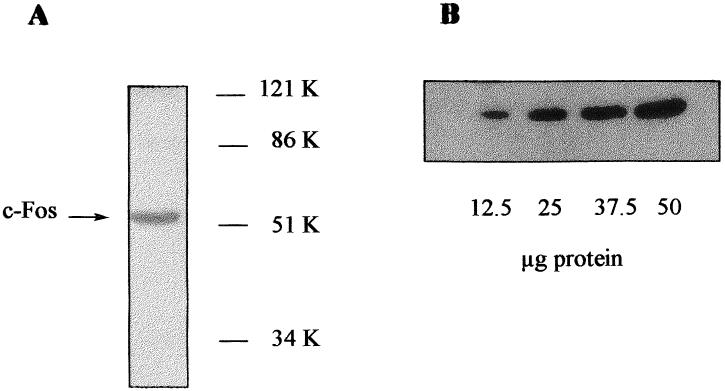
Fig 1.

Western blot analysis of c-Fos in mice liver homogenates. (A) Liver homogenate (50 μg of protein) was loaded onto 10% polyacrylamide gel. After transfer, the membrane was incubated with the primary antibody (directed against a N-terminal fragment containing residues 75–155 of v-Fos but conserved both in human and mouse c-Fos). After washing, the membrane was incubated with a secondary antibody and then developed with the ECL system. (B) 10% Polyacrylamide gel was loaded with increasing amounts of liver homogenate protein and processed as indicated above.
