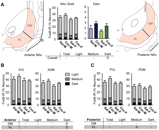
Figure 7. Cocaine selectively increased the proportions of neurons darkly stained for FosB in the nucleus accumbens (NAc) Shell of male and female rats.
A) A Bar graphs showing the percentage of all neurons in saline- (Sal) and cocaine- (Coc) treated male (M) and female (F) rats that were FosB-IR. Each bar shows the percentage of all neurons in each treatment group that were light, medium, or darkly stained (middle left, NAc Shell), and darkly stained cells only (middle right, Dark). The partitioning scheme used to define dorsomedial (DM) and ventrolateral (VL) zones for anterior (left) and posterior (right) NAc Shell are also shown. B and C) Bar graphs showing the proportion of FosB-IR cells in each anatomical zone of the anterior (B) and posterior (C) NAc Shell. Each bar shows the percentage of all neurons in each treatment group that were light, medium, or darkly stained. The tables summarize statistically significant differences (p<0.05) for the NAc Shell as a whole (A), in the anterior (B) and the posterior (C) anatomical zones. “D” indicates a statistically significant drug effect, and “S” indicates a significant sex difference (See text).

