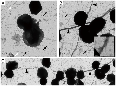
Figure 4. NafA regulates bacterial piliation.
Piliation of the wild-type strain (A) and the ΔNafA strain (B, C) was analyzed by transmission electron microscope after negative staining. White arrows mark membrane blebs and black arrows mark pili. Arrowheads in panel B and C indicate thick bundled pilus structures. The inset in panel B shows a higher magnification of the boxed area, which displays the bundled pili. An overview of the ΔNafA strain piliation is shown in panel C. Black scale bars show 0.5 µm and white scale bars 50 nm.

