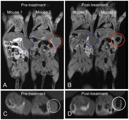
Figure 7. T2-weighted MR images of nude mice with breast tumor obtained (A) before and (B) after injection of MR contrast agent, obtained using a 3 Tesla MR scanner.
Mouse 2 was injected with Mag@SiO2 nanoparticles as T2 contrast agent, while Mouse 1 was injected with saline as a control. Tumor sites in the control (mouse 1) and in the treated mouse (mouse 2) have been labelled as blue and red circles respectively. Panels C and D show the higher magnification transverse section images of tumor site corresponding to Panels A and B respectively, wherein tumor region injected with MR contrast agent has been highlighted using white circles.

