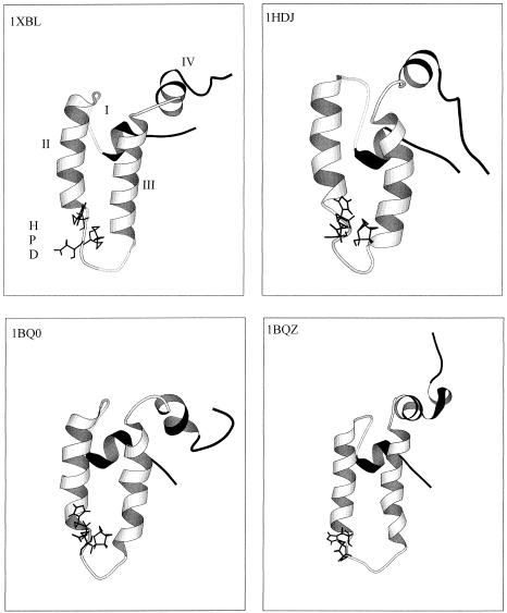
Fig 1.
Ribbon representation of the structures of the DnaJ and HDJ1 J domains as determined using nuclear magnetic resonance. The PDB codes are 1XBL (Pellechia et al 1996) and 1BQ0 and 1BQZ (Huang et al 1999) for the J domain from E coli DnaJ and 1HDJ (Qian et al 1996) for the J domain from human HDJ1. The structures were visualized in Molscript (Kraulis 1991). The HPD motif is indicated as sticks. Helices II and III are in white, with the more mobile helices I and IV in black. The helices and HPD motif are labeled on the 1XBL structure

