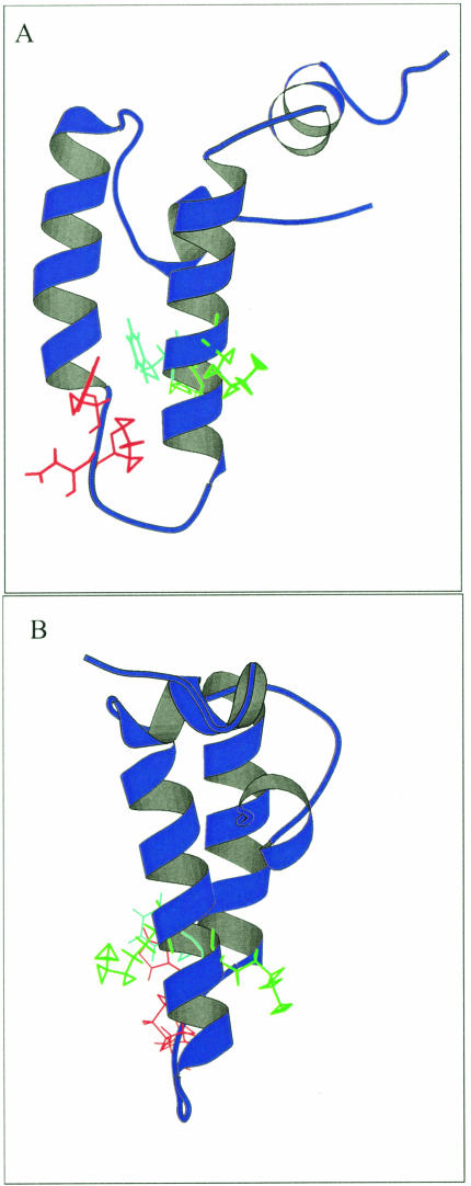
Fig 5.
Ribbon representation of the J domain in E coli DnaJ showing the KFK motif and the HPD motif. The structure used is 1XBL. The HPD motif is in red, the phenylalanine is in light blue, and the 2 lysines on either side of the phenylalanine are in green. The model was made using Molscript (Kraulis 1991). (A) Visualization of the phenylalanine residue with respect to the HPD motif. (B) Visualization of the orientation of the flanking lysine residues

