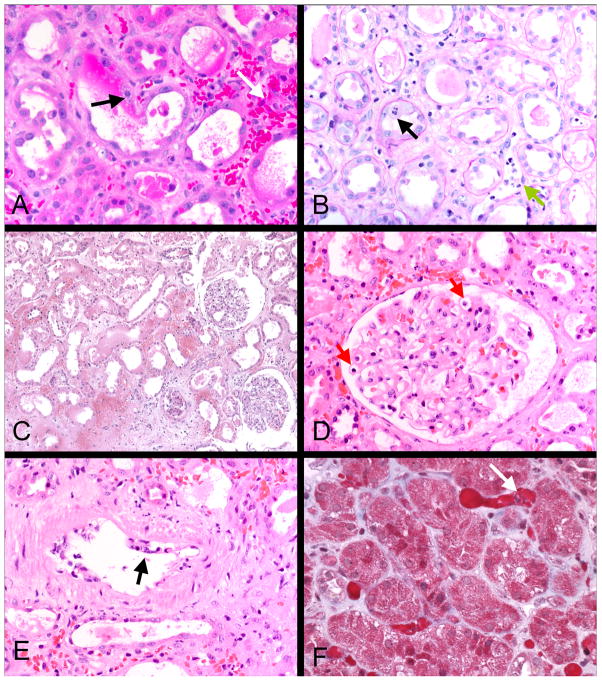
Figure 3.
Light microscopy of representative engraftment syndrome cases: Interstitial hemorrhage and congestion are prominent in these cases, as illustrated in A, C and F. Intracapillary mononuclear and polymorphonuclear cells are present in peritubular and glomerular capillaries (B green arrow and D red arrows), a hallmark of this condition. Tubular injury is also characteristically present, as shown by loss of brush border and tubular epithelial mitoses (black arrow B). Two cases had transient arterial endothelial inflammation, as shown in E (arrow); some cells were neutrophils, in contrast to the usual T cell mediated rejection. Little or no interstitial inflammation or tubulitis was evident in contrast to cell mediated rejection. A, C, D, and E, H&E; B, PAS; F, trichrome.

