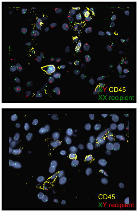
Figure 9.
Fluorescence in situ hybridization (FISH) of ES biopsies to determine the origin of the leukocytes in the capillaries. All cells stain for recipient type DNA. In the upper panel the donor was male and recipient female. All of the CD45+ cells stain for XX and none for XY. CD45− cells are largely donor type (tubules, endothelium). In the lower panel, the donor was female and recipient male, and all the CD45+ cells are positive for XY, and none for XX. CD45 (yellow), X chromosome (green), Y chromosome (red) and DAPI nuclear stain (blue).

