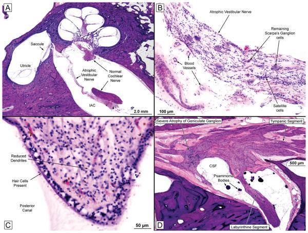
FIG. 1.
A, Low-power view of the left ear (near-mid modiolar section) showing an atrophic vestibular nerve and a normal-appearing cochlear nerve. The cochlear turns, saccule, and utricle seemed normal.
B, High-power view of the left vestibular nerve within the internal auditory canal. The nerve was severely atrophic, but there was no evidence of inflammation or vasculitis. There was severe loss of Scarpa's ganglion cells.
C, Crista of the left posterior semicircular canal showing a good population of hair cells and supporting cells in the neuroepithelium. There was a reduction of dendrites within the stroma.
D, Facial nerve from the right ear. There was severe diminution of the number of geniculate ganglion cells. CSF indicates cerebrospinal fluid.

