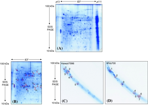
Fig. 2.
Overview of the cell wall proteome analyzed from the mature tomato stem and separated in two as well as in three gel dimensions. (A) The resolution of proteome with 2-D IEF/SDS–PAGE in pI 3–11 non linear and 100–10 kDa molecular mass. The poorly resolved proteins on the basic pI range were seen as vertical streaking. (B) A representative 2-D IEF/SDS gel resolved in the same pI 3–11 non linear and 100–10 kDa molecular mass range but stained after cutting out the gel region with the poorly resolved vertical streak. The poorly resolved basic proteins without fixing and staining served as the starting material for the 3rd SDS–PAGE. Twenty spots were randomly picked out to check the presence of secretion signals of the cell wall proteins extracted with the method applied. The identity of each encircled spots and the information of secretion signals are given in Table 5. (C) and (D) 3rd dimension SDS gels showing the resolution of the unresolved vertical streak. The streaked gel piece was cut out before fixing as well as staining and separated again by SDS–PAGE. The encircled spots in (C) and (D) were differentially expressed in resistant (Hawaii7996) and susceptible (WVa700) species after pathogen inoculation, however in two replicates. The identity of all proteins was given in Table 4 with the spot numbers corresponds to each other

