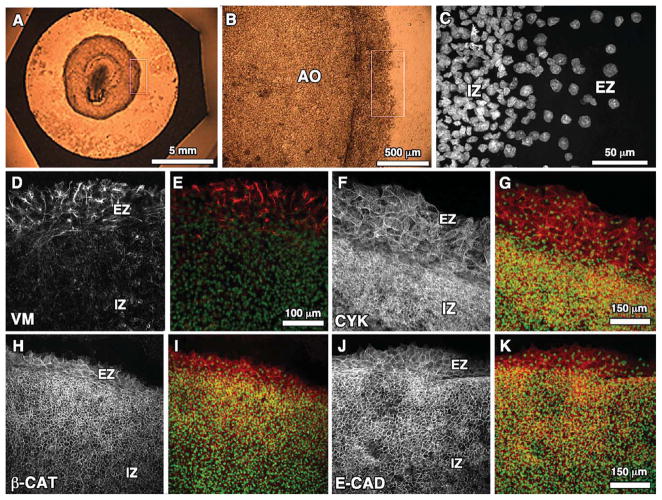
Fig. 1.
A–C: Progressive zoom-ins of the blastoderm edge zone of the quail embryo indicated by the white box in A. C: Sytox-labeled nuclei for region outlined in B. D, F, H, J: Representative wide-field (20×) confocal immunoflourescent images for vimentin (VM), cytokeratin (CYK), β-catenin (β-cat), and E-cadherin (E-cad), respectively, at the edge zone (EZ) and innner zone (IZ) of Stage-4 quail blastoderm. E, G, I, K: Each marker is shown merged with Sytox nuclear label.

