Fig. 2.
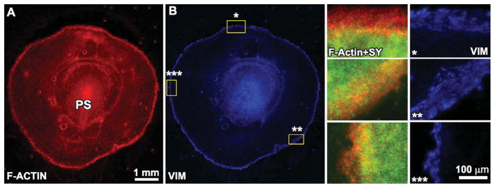
A,B: A wide-field (2.5×) montage of F-actin (rhodamine-phalloidin) and vimentin immunofluorescence for a stage-4 quail embryo. The starred regions in B at different regions around the embryo perimeter are shown at higher resolution on the right side. Note the variability in the width of the edge zone in these different regions, which is made clear by the edge-restricted vimentin expression. PS, primitive streak.

