
Figure 3.
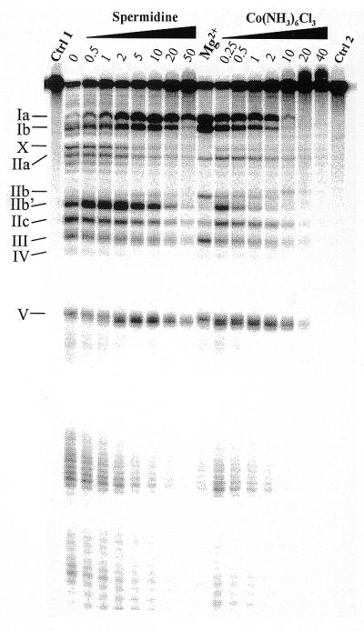
Lead(II)-induced cleavage patterns of M1 RNA in the presence of increasing concentrations (in mM) of the Mg(H2O)62+ analog Co(NH3)63+ and spermidine, as indicated. Cleavage was performed at the indicated concentrations and 37°C, as outlined in Materials and Methods and Figure 2 legend, with the exception that the time of incubation in the presence of Pb2+ was 7 instead of 6 min. Ctrl 1, incubation in the presence of 50 mM Tris–HCl pH 7.5, 100 mM NH4Cl, 10 mM Co(NH3)63+, 10 mM spermidine, 10 mM MgCl2 and without Pb2+; Ctrl 2, as Ctrl 1 but incubation in the presence of 50 mM Tris–HCl pH 7.5 and 100 mM NH4Cl (without Pb2+) only; Mg2+, Pb2+ cleavage in the presence of 50 mM Tris–HCl pH 7.5, 100 mM NH4Cl and 5 mM MgCl2. Roman numerals indicate the cleavage sites as shown in Figure 1.