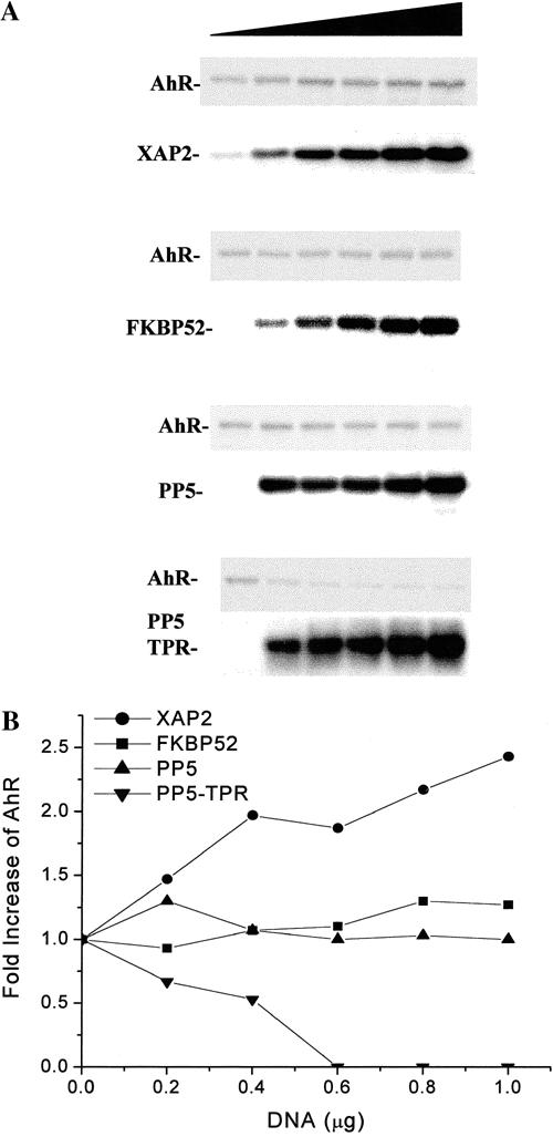
Fig 2.

XAP2 specifically enhances the level of AhR in COS-1 cells when compared to other hsp90-binding TPR-containing proteins. COS-1 cells were transfected in 6 well dishes with 1 μg of pcDNA3/βmAhR and 0, 0.2, 0.4, 0.6, 0.8, or 1.0 μg of pCI/XAP2, pCI/FKBP52, pCMV6/PP5-FLAG, or pCMV6/PP5-TPR-FLAG (shaded triangle) and brought to a total of 2 μg vector/dish with pCI vector. (A) Total cell lysates were isolated, and 75 μg of each lysate were resolved by SDS-PAGE, transferred to PVDF membrane, and analyzed by immunoblot analysis. XAP2 and FKBP52 were detected with XAP2 polyclonal and FKBP52 monoclonal antibodies. PP5-TPR and PP5-TPR-FLAG were detected with anti-FLAG M2 antibody. AhR was detected with the anti-AhR monoclonal antibody RPT1. XAP2 was visualized with [125I]-DAR, and the AhR, FKBP52, PP5-FLAG, and PP5-TPR-FLAG were visualized with [125I]-SAM. (B) The graph depicts the fold change in AhR levels obtained in the presence of TPR-containing proteins after phosphorimaging of the blots. This experiment has been repeated 3 times with essentially the same results
