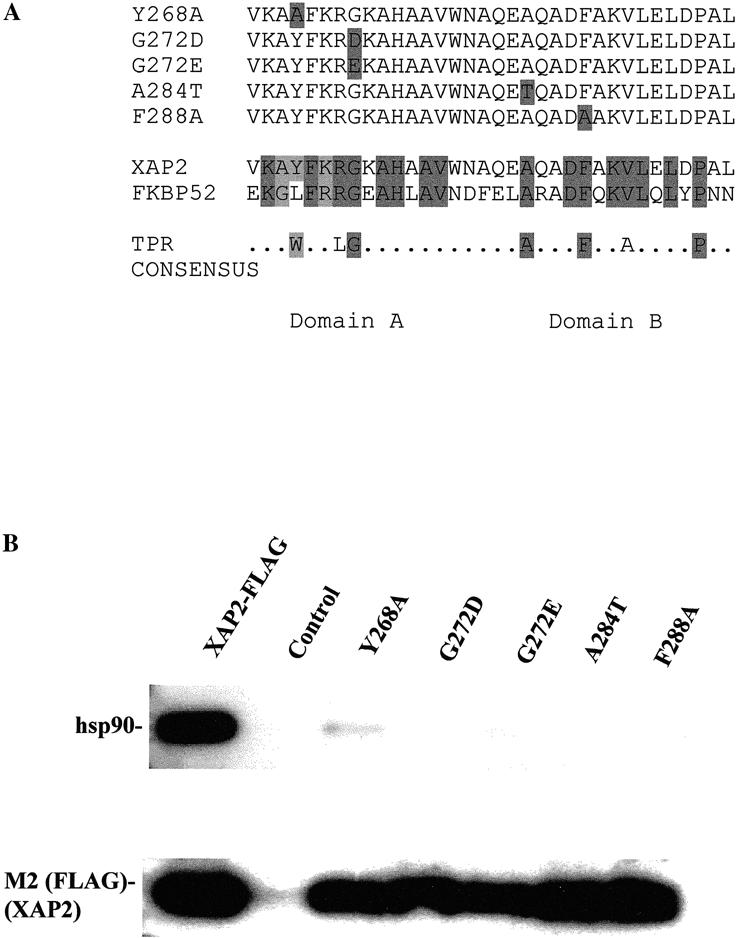
Fig 3.

Schematic representation of XAP2 TPR mutants and their ability to bind to hsp90 in COS1 cells. (A) Top panel: Black boxes indicate amino acid substitutions in the respective mutant. Bottom panel: Black boxes indicate conserved amino acid residues between XAP2 and FKBP52. The TPR consensus was generated from CDC16, CDC23, CDC27, SSN6, and SK13 (Lamb et al 1995). (B) COS-1 cells were transiently transfected with pCI/XAP2-FLAG, pCI/XAP2-FLAG-TPR mutants, or pCI (control), and cell lysate was isolated, immunoabsorbed with the M2 anti-FLAG affinity resin, eluted with FLAG peptide, resolved by SDS-PAGE, and transferred to PVDF membrane, followed by immunoblot analysis. Hsp90 was visualized with polyclonal antibodies raised against hsp84/86 and [125I]-DAR, and XAP2-FLAG was visualized with anti-FLAG M2 antibody and [125I]-SAM
