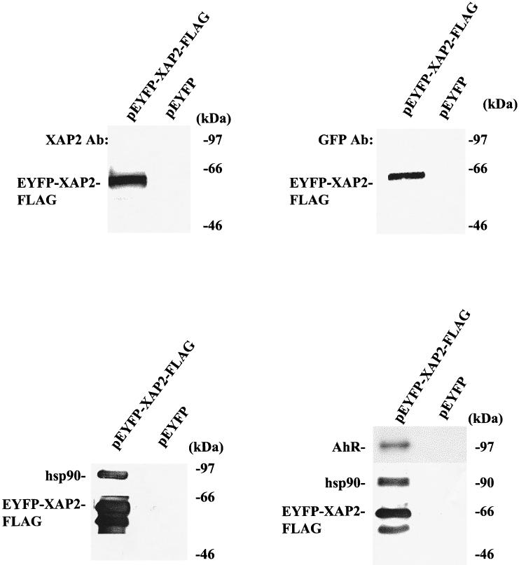
Fig 7.

EYFP-XAP2 binds to hsp90 and to AhR/hsp90 complexes in COS-1 cells. Upper panels: pEYFP-XAP2-FLAG or pEYFP alone was transiently transfected in COS-1 cells, cytosol was isolated, and 150 μg of protein were resolved by SDS-PAGE and transferred to PVDF, followed by immunoblot analysis with XAP2 polyclonal antibodies or anti-GFP monoclonal antibodies. Antibodies were visualized with DAR-P and GAM-P, respectively. Lower panels: pEYFP-XAP2-FLAG or pEYFP were transiently transfected in COS-1 cells (left panel); pEYFP-XAP2-FLAG or pEYFP were transiently cotransfected with pcDNA3/βmAhR in COS-1 cells (right panel). In both experiments, cytosol was isolated and immunoabsorbed with the M2 affinity resin, and complexes were eluted with FLAG peptide and resolved by SDS-PAGE, transferred to PVDF membrane, and analyzed by immunoblot analysis. pEYFP-XAP2-FLAG was visualized with the M2 antibody, AhR with the RPT1 antibody, and hsp90 with rabbit polyclonal antibodies against hsp84 and hsp86. M2 antibody was visualized with GAM-P, hsp90 with DAR-P, and AhR with GAM-P by ECL
