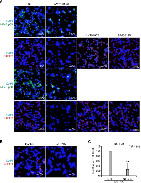
Figure 4.
(A) B cells cultured with BAY1170-82 (10 LY294002 (1 or SP900125 (10 for 24h were examined bv immunofluorescence staining of BAFF-R, NF-p65 and NF-p50. (B and C) B cells treated with shRNAs(2 g/ml) to NF-κB or GFP(negative control) were analyzed by confocal microscopy (B) and real-time PCR (C). *P < 0.01.

