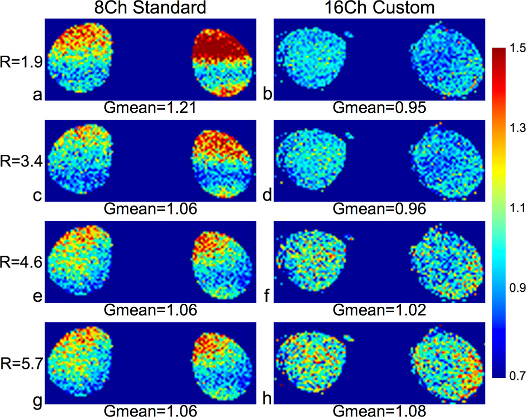
Figure 3.
G-Factor maps of a coronal slice (reformatted from a 3D acquisition) of a healthy volunteer with L/R and S/I acceleration using the 8-channel standard coil (column 1) and the 16-channel custom coil (column 2). We acquired the full k-space data with a 3D SPGR sequence and applied ARC acceleration in post-processing – 2× S/I and 1×, 2×, 3×, or 4× L/R for effective accelerations of R = 1.9, 3.4, 4.6 and 5.7. We calculated ARC g-factors using the pseudo multiple replica method. Both coils had fairly low g-factors for all ARC accelerated images, but the custom coil had lower mean and peak values for R = 1.9, 3.4, and 4.6.

