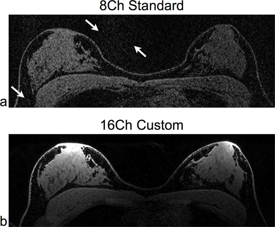
Figure 6.
Accelerated (R = 3.9) 3D SPGR Scans of a normal volunteer using identical parameters for both coils: a 3D SPGR sequence, spectral-spatial fat suppression, and 4× ARC acceleration in the L/R direction (R = 3.9). We compared the 8-channel standard coil (a) and the 16-channel custom coil (b). There is a substantial improvement in signal quality in the 16-channel custom coil image, and there is little or no visible parallel imaging noise. The white arrows in (a) highlight the parallel imaging noise with the 8-channel standard coil. For the 16-channel custom coil image, the low sensitivity at the heart reduces cardiac motion artifact. We windowed both images to the same level and acquired both scans with identical parameters: axial, 1:13 scan time, 512×272×312 matrix size, and 0.7×1.3×1.0 mm resolution.

