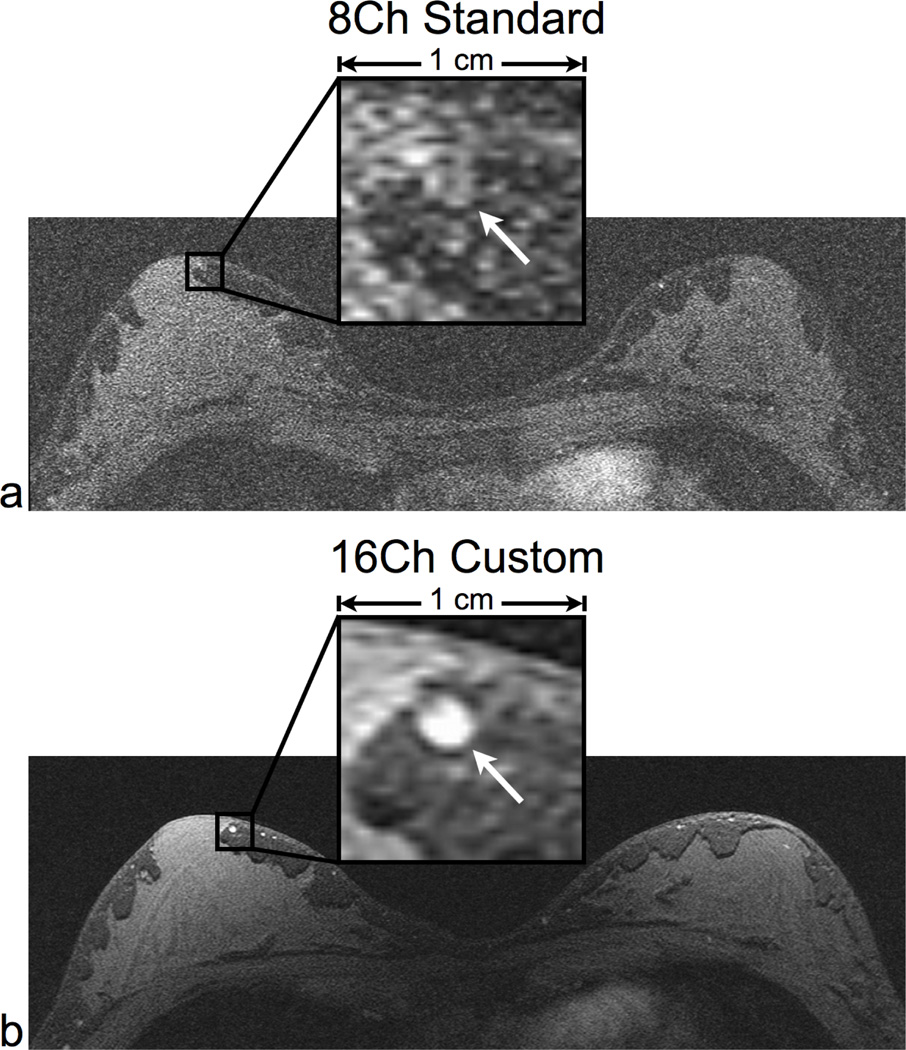
Figure 8.
High spatial resolution axial scans in a normal volunteer using the same parameters for both coils: a 3D SPGR sequence with no parallel imaging. We compared the 8-channel standard coil (a) and the 16-channel custom coil (b). The 16-channel coil image clearly delineates fine morphological features such as a 2 mm wide blood vessel (see arrow). The low SNR in the 8-channel coil image limits the ability to identify these features. The scan parameters were: 2:43 scan time, 1024×512×32 matrix size, and 0.3×0.6×0.6 mm resolution.

