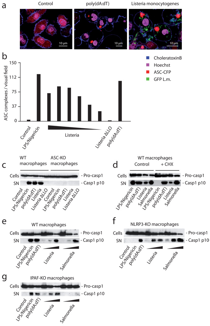
Figure 1. Listeria triggers caspase-1 activation through ASC, yet is unimpaired in cells solely deficient in NLRP3 or IPAF.
(A) Wild type macrophages stably expressing ASC-CFP were treated with L. monocytogenes expressing GFP, transfected with poly(dA:dT) or left untreated. 6 hours after stimulation, the formation of ASC complexes was quantified using confocal microscopy. (B) ASC-CFP expressing macrophages were treated with ATP/LPS, decreasing amounts of L. monocytogenes (MOI 20 – 0.625 in steps of 50%), a mutant L. monocytogenes strain lacking LLO (Listeria ΔLLO, MOI 20) or transfected with poly(dA:dT) and formation of ASC complexes was quantified using epifluorescence microscopy. (C) Wild type macrophages or ASC-deficient macrophages were treated with the stimuli as above (L. monocytogenes MOI of 10). 6 hours after stimulation pro-caspase-1 (pro-casp1) expression was assessed in cell lysates (cells), while caspase-1 activation (casp1 p10) was assessed in the supernatant (SN) using western blot. (D) Wild type macrophages were treated with 20 ng /ml cycloheximide or left untreated. After 30 min cells were stimulated as indicated (L. monocytogenes and S. enterica MOI of 10) and caspase-1 expression and activation was assessed as above. (E) Wild type, (F) NLRP3- or (G) IPAF-deficient macrophages were stimulated as indicated (L. monocytogenes / S. enterica MOI of 50, 10 or 1) and 6 hours after stimulation caspase-1 expression and activation was assessed. One representative experiment out of three (A, B, D) or four (C, E, F, G) independent experiments is depicted.

