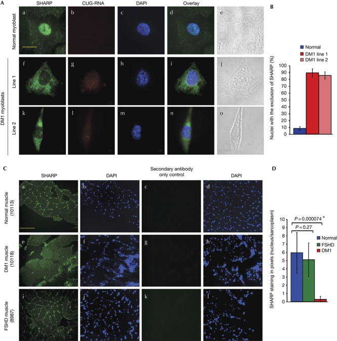
Figure 3.
SHARP is abnormally distributed in DM1. (A) Endogenous SHARP is shown in green (a,f,k). Expanded CUG tracts were detected by fluorescence in situ hybridization (red; g,l). Nuclear DAPI staining of normal and DM1 myoblasts (c,h,m) are shown. Merged images demonstrate that endogenous SHARP localizes primarily in the nucleus in normal myoblasts (d) and predominantly in the cytoplasm in the two DM1 myoblast lines (i,n). Bright-field images (e,j,o) are shown. Scale bar, 15 μM. (B) Graphical representation of nuclei with exclusion of SHARP in 90 normal and DM1 cells. Error bars (±) represent standard deviation (n=3). (C) Cross-sections of normal human (10113), DM1 (10118) and FSHD (8987) muscles were immunostained with SHARP antibodies followed by DAPI staining and visualized using × 20 magnification. Endogenous SHARP is shown in green (a,e,i). A staining control in which only the secondary antibody was used is shown in panels c,g,k. Nuclei are stained with DAPI (b,f,j,d,h,l). Scale bar, 175 μm. (D) Graphical representation of nuclear/sarcoplasmic SHARP staining in pixels of normal, FSHD and DM1 muscle is shown. Error bars (±) represent standard deviation (n=8). P-values were determined by paired Student's t-test. Quantification method is described in supplementary Fig S4C online. DAPI, 4,6-diamidino-2-phenylindole; DM1, myotonic dystrophy; FSHD, facioscapulohumeral muscular dystrophy; SHARP, SMART/HDAC1-associated repressor protein.

