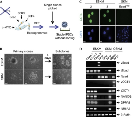
Figure 3.
E-cadherin can replace OCT4 in reprogramming. (A) Derivation of ESKM-induced pluripotent stem cell clones following viral transduction of MEFs in the presence of Ecad but the absence of OCT4. Transduced MEFs were seeded and embryonic-stem-cell-like colonies were observed. (B) Morphology of two independent primary clones photographed 12 days after viral transduction of MEFs with ESKM (upper panel) or SKM (lower panel). Primary clones were cultured for 4 days on feeder cells. Morphology of ESKM or SKM cells is shown in the right panels. Magnification × 400 (primary clones on feeder cells, two left panels); scale bars, 50 μm and × 100 (subclones, right panel); scale bars, 200 μm. (C) Immunofluorescence analysis of established ESKM (2, 3) and SKM cells for expression of OCT4. Nuclei were stained with DAPI. Magnification × 400; scale bars, 50 μm. (D) Reverse transcription–polymerase chain reaction analysis was performed with three independently generated ESKM clones (1, 2 and 3), ESKM-infected MEFs 2 days post-infection (2d pi), SKM subcloned cells (SKM), SKM-infected MEFs, OSKM-derived iPSCs and OSKM-infected MEFs. Expression levels of Ecad (v/t), Ncad, OCT4 (v/t), NANOG, DPPA5 and NR5A2 of cells were determined. DAPI, 4,6-diamidino-2-phenylindole; ESKM, pMXs-Ecad retrovirus in combination with SOX2, KLF4 and c-MYC; iPSC, induced pluripotent stem cell; MEF, mouse embryonic fibroblast; MET, mesenchymal-to-epithelial transition; OSKM, OCT4, SOX2, KLF4 and c-MYC.

