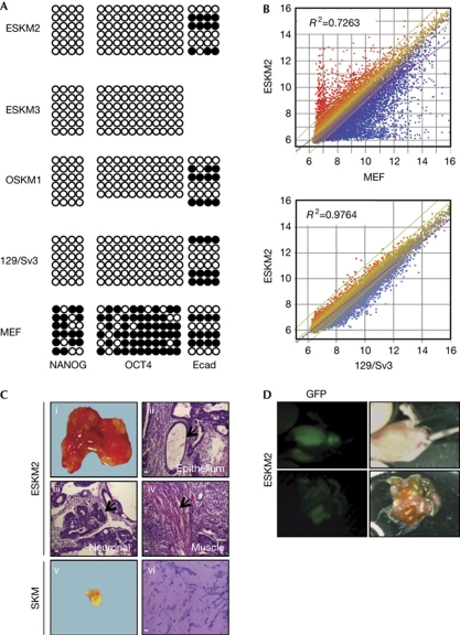
Figure 4.
ESKM induced pluripotent stem cells fulfil all criteria of pluripotent stem cells. (A) Bisulphite sequencing of promoter regions of NANOG, OCT4 and Ecad, of ESKM cells (clone 2 and 3), OSKM cells (clone 1), mouse embryonic stem cells (mESCs; 129/Sv3) and mouse embryonic fibroblasts (MEFs). Shown are 5–6 reactions for each region. (B) Global gene expression profiling of ESKM-iPSCs (clone 2) in comparison with MEFs and mESCs. Similarity is shown by the R2 value. Three biological replicates of each cell line were included for analysis. (C) Whole morphology of tumours derived from ESKM-derived iPSCs (ESKM 2; 1) or SKM-derived cells (v). Histological analysis of tissue sections by haematoxylin and eosin staining of differentiated tumours (ESKM2) comprising epithelial (ii), neuronal (iii) and muscle-derived (iv) structures, or of a undifferentiated tumour (SKM; vi). Representative tumours are shown out of n=3 injections/cell clone, compare supplementary Table 3 for further details. Magnification of haematoxylin and eosin-stained sections × 25; scale bars, 500 μm. (D) Localization of green fluorescent protein (GFP)-positive areas in offspring following injection of stable GFP-expressing ESKM-iPSCs (clone 2) in blastocysts. ESKM, pMXs-Ecad retrovirus in combination with SOX2, KLF4 and c-MYC; OSKM, OCT4, SOX2, KLF4 and c-MYC.

