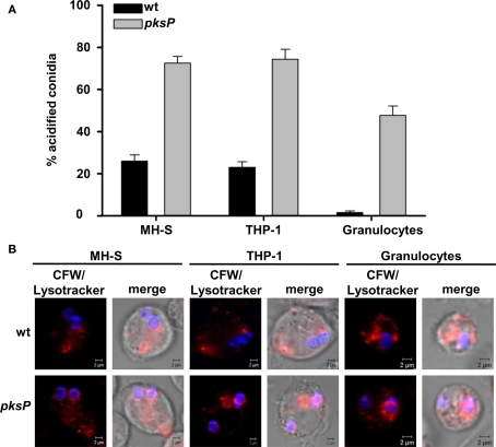
Figure 1.
Detection of conidia in acidified compartments after phagocytosis by MH-S and THP-1 cells, and neutrophil granulocytes. (A) Percentages of conidia present in acidic phagolysosomes. Calcofluor white (CFW)-labeled conidia were intracellularly colocalized with LysoTracker Red-DND99. Data represent the mean values and SD of three experiments. (B) Representative micrographs showing colocalization of conidia stained using CFW (blue) with acidified compartments visualized by LysoTracker labeling (red). Size bar, 2 μm.

