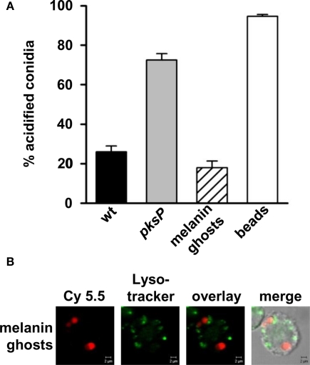
Figure 3.
Influence of A. fumigatus wild-type-derived melanin ghosts on phagolysosomal acidification. (A) Percentage of intracellular conidia residing in acidified phagolysosomes after ingestion by MH-S cells. Latex beads were used as a positive control. Values represent the mean + SD of three experiments. (B) Representative micrographs showing colocalization of melanin ghosts, stained prior to phagocytosis with biotinylated anti-melanin camelid antibody and Cy5.5 conjugated streptavidin (red), with acidified compartments visualized by LysoTracker labeling (green). Size bar, 2 μm.

