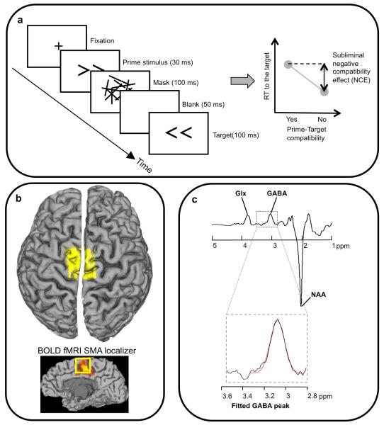
Figure 1. Methodology for masked priming and GABA spectroscopy.
a, target arrows were preceded by masked primes presented below the threshold for conscious discrimination. For the stimulus timing illustrated, responses tend to be slower when prime and target are the same (compatible) than when they are not (right hand illustration). This is the measure of subliminal suppression, and the magnitude differs between individuals. b, the MRS voxel (yellow, (3 cm)3 voxel) was placed over the anatomical location of SMA. As a check on voxel placement, for two participants we acquired a functional localiser for the SMA using fMRI (see Methods and bottom sagital view). Edited MR spectra (c) allow the quantification of GABA concentration by extracting the area under the GABA peak [6, 8, 9, 48] (glutamine/ glutamate, Glx, and N-acetyl-aspartate, NAA, peaks are also marked). The peak will also contain co-edited macromolecules. See Methods and supplementary fig S2 for more details and individual spectra.

