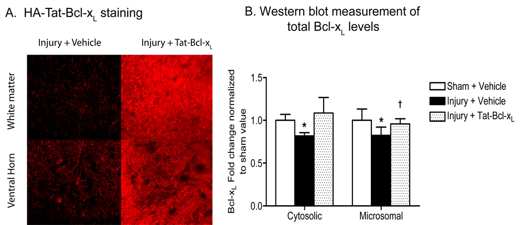
Fig. 1. Protein transduction of Tat-Bcl-xL in the injured spinal cord 24h after trauma.
A. Immunofluorescence staining of a transverse section of spinal cord 3mm rostral to the lesion epicenter, 24h after injury, using an antibody against HA-tag. The HA immunoreactivity was strong in gray and white matter in Tat-Bcl-xL-treated, but not vehicle-treated spinal cords. Scale bar: 50 µm. B. Quantitation of cytosolic and microsomal Bcl-xL levels based on western blot analyses. We used an antibody against a sequence surrounding Asp61 in the loop region of Bcl-xL that recognized both endogenous Bcl-xL and Tat-Bcl-xL. Total Bcl-xL levels at cytosolic and microsomal fractions increased in Tat-Bcl-xL-treated animals to levels comparable to those of sham-treated cords. Bcl-xL expression values were normalized to sham values and presented as mean ± SD. n=4/5 per group as indicated in Table 1. * p<0.05 compared to sham-vehicle group; † p<0.05 compared to the injury-vehicle treated group; Two way ANOVA with Tukey’s post-hoc correction).

