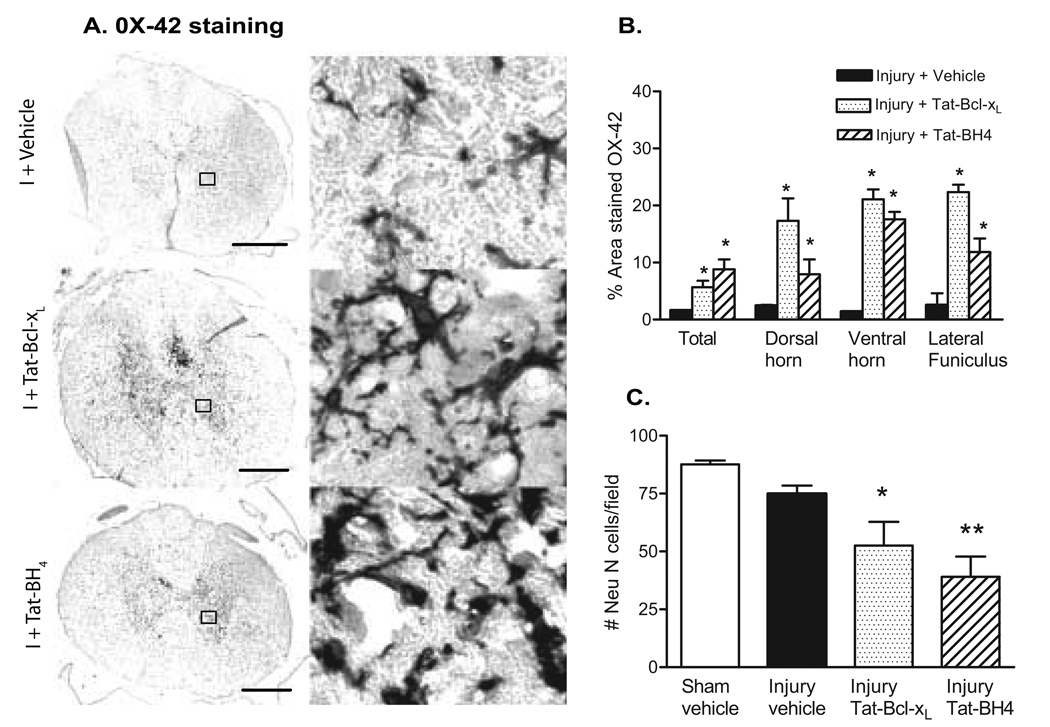
Fig. 5. Neuronal and microglial/macrophage densities after Tat-Bcl-xL and Tat-BH4 treatment.
A. Semi-quantitative analysis of OX-42 staining 4mm rostral to lesion epicenter in Tat-Bcl-xL- (n=3); Tat-BH4- (n=3) and vehicle-treated rats (n=3) 7 days after SCI. Data represent the proportional area of OX-42 staining in a 6.25mm2 region (total OX-42 labeling) or a 0.0625mm2 region at the dorsal horn, ventral horn and lateral funiculus. (See methods for details of quantification methods). B. Representative example of OX-42 staining of microglia and macrophages 4mm rostral to the lesion epicenter, 7 days after injury. Left panel: cross sectional OX-42 staining showing increased microglial/macrophage activation in Tat-Bcl-xL- and Tat-BH4- treated injured spinal cords in comparison to vehicle-treated injured spinal cords. Scale bar: 350µm Right panel: high magnification of the area marked in the left panel as squares, shows intense labeling of OX-42 in long processes and round cell bodies surrounding gray matter cells, likely neurons, in Tat-Bcl-xL- and Tat-BH4-treated injured spinal cords, and a moderate OX-42 labeling in vehicle-treated injured spinal cords. C. Quantitative analysis of NeuN-labeled cells in the dorsal and ventral horn. Neurons were counted 4mm rostral to the lesion epicenter in sections of Tat-Bcl-xL(n=3), Tat-BH4 (n=3) and vehicle-treated rats (n=3) 7 days after SCI. The number of Neu-N-positive cells was significantly lower in the Tat-Bcl-xL- and Tat-BH4-treated injured spinal cords in cord (*p<0.05). Sections are adjacent to those used for OX-42 analysis.

