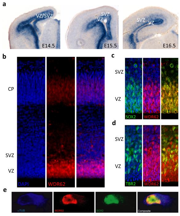
Fig 4. Wdr62 expression in the developing mouse brain.
a, Wdr62 expression is enriched in the VZ and SVZ as seen with in-situ hybridization. b, WDR62 protein(red) distribution reveals a similar pattern. c, d, WDR62 (red) localizes to the nuclei and is expressed by neural stem cells and intermediate progenitors, as marked by SOX2 and TBR2 expression (green), respectively. e, Immunofluorescent staining for α-tubulin (cytoplasmic, blue), S0X2 (nuclear, green) and WDR62 (red) in E12.5 cortical neural progenitor cells reveals that the distribution of the WDR62 overlaps with that of SOX2 and is predominantly nuclear. (Nuclear staining by DAPI (blue) in b-d; Right most panels are composite images in b-e).

