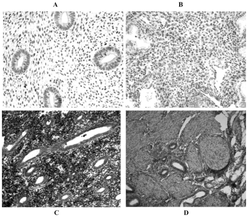
FIGURE 1.

TF immunohistochemistry. (A) Normal proliferative endometrium showing low to no TF staining in glands or stromal cells. (B) Normal secretory endometrium showing decidualized stromal cell staining. (C) Ectopic endometriotic implant from proliferative phase endometrium with glandular staining. (D) Eutopic late proliferative phase endometrium from patients with endometriosis with glandular staining (×20).
