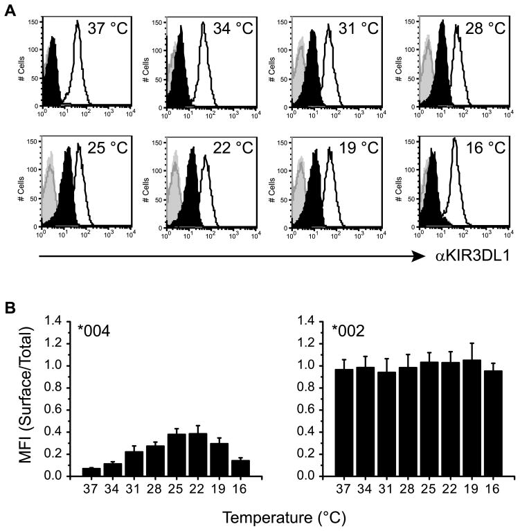
Figure 1. Culture at lower-than-physiological temperatures increases cell surface expression of KIR3DL1*004.
A, NKL cells transfected to express KIR3DL1*002-GFP (line histograms) or KIR3DL1*004-GFP (filled histograms) were incubated for 16 h at the temperatures indicated (top right of histogram). Cell surface expression of KIR3DL1 was detected by flow cytometry after staining with anti-KIR3DL1 mAb 177407 (black) or an isotype-matched control (grey). B, Mean fluorescence intensity (MFI) ratios for histograms shown in A, are plotted as surface (mAb)/total (GFP) KIR3DL1. Mean and SD across 3 independent experiments are shown. Temperature induced changes in KIR3DL1*004 but not KIR3DL1*002 expression are statistically significant as assessed by one-way ANOVA (P < 0.0001 and P = 0.2054 respectively). For KIR3DL1*004 expression, post test analysis for linear trend at temperatures permitting exocytosis (37–22 °C) is also statistically significant (P < 0.0001).

