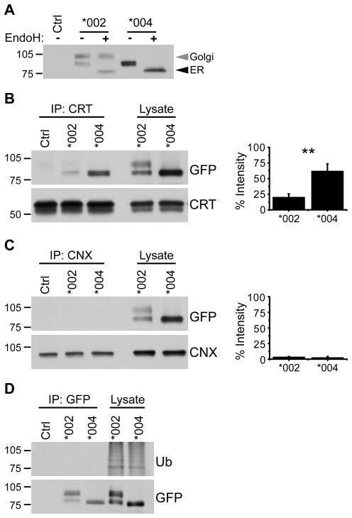
Figure 3. KIR3DL1*004 is retained in the ER.
A, KIR3DL1 was immunoprecipitated from lysate of NKL cells expressing KIR3DL1*002-GFP (*002) or KIR3DL1*004-GFP (*004) using anti-GFP antibody. Immunoprecipitated protein was treated with Endoglycosidase H (Endo H) and KIR3DL1 was detected by Western blotting for GFP. Ctrl = NKL transfected to express KIR3DL1*002 lacking GFP. Endo H-resistant (Golgi) and -sensitive (ER) species are marked (grey and black arrowheads respectively). B, Calreticulin (CRT) or C, Calnexin (CNX) was immunoprecipitated from NKL transfectants. Ctrl = NKL transfected to express KIR3DL1*002 lacking GFP. Co-immunoprecipitated KIR3DL1 was detected by Western blotting for GFP (upper). Membranes were stripped and re-probed for the immunoprecipitated chaperone protein (lower). Graphs show quantification of Western blots (right). Band intensities are plotted with co-immunoprecipitated KIR3DL1 expressed as a % of signal from lysate. Mean and SD across 3 independent experiments are shown. Statistical significance was assessed by unpaired Student’s t test. ** P = 0.0062. D, KIR3DL1 was immunoprecipitated as in A followed by Western blotting for ubiquitin (upper). Membrane was stripped and re-probed for GFP (lower). The migration position of molecular weight markers (kD) is shown at the left of each panel.

