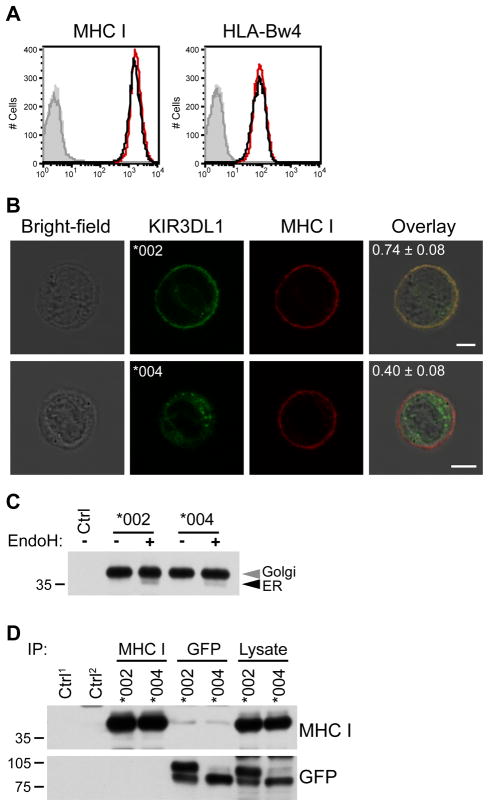
Figure 5. KIR3DL1*004 does not retain MHC class I inside the cell.
A, Cell surface expression of MHC class I protein was analyzed by flow cytometry. NKL cells transfected to express KIR3DL1*004-GFP (black) or KIR3DL1*002-GFP (red) were stained with mAb against all MHC class I (left) or HLA-Bw4 (right). Isotype-matched controls are shown in grey (KIR3DL1*004 transfectants = line, KIR3DL1*002 transfectants = fill). B, NKL expressing GFP-tagged KIR3DL1 (green, second column) were fixed and stained for MHC class I (red, third column). Fluorescence/bright-field overlays are also shown (fourth column) and overlap of red and green signals (yellow) was quantified with Pearson’s correlation coefficient (mean ± SD shown in panel). 62 and 78 cells were imaged for KIR3DL1*002 (*002) and KIR3DL1*004 (*004) respectively. Scale bars = 5 μm. C, MHC class I was immunoprecipitated from NKL transfectants. Ctrl = immunoprecipitation with isotype-matched control in KIR3DL1*002-GFP transfectants. Immunoprecipitated protein was treated with Endoglycosidase H (Endo H) and detected by Western blotting for MHC class I. D, Co-immunoprecipitation of GFP-tagged KIR3DL1 and MHC class I was assessed in NKL transfectants. Immunoprecipitation using GFP and MHC class I-specific antibodies (top) was followed by Western blotting for the reciprocal protein (right). Endo H-resistant (Golgi) and -sensitive (ER) species are marked (grey and black arrowheads respectively). Ctrl1 = isotype-matched control immunoprecipitation from NKL transfected to express KIR3DL1*002-GFP. Ctrl2 = anti-GFP immunoprecipitation from NKL transfectants expressing KIR3DL1*002 which lack GFP. The migration position of molecular weight markers (kD) is shown at the left of panels in C and D.

