
Fig 3.
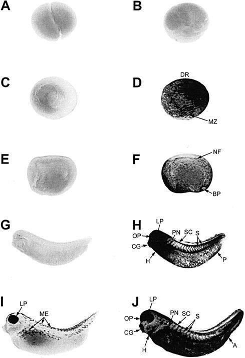
Spatial pattern of hsp70 mRNA accumulation during early Xenopus laevis development. Whole-mount, in situ hybridization with digoxygenin-labeled hsp70 antisense riboprobe was performed with control (22°C for 1 hour; panels A, C, E, G, and I) and heat shocked (33°C for 1 hour; panels B, D, F, H, and J) X laevis embryos at cleavage (stage 3; panels A and B), gastrula (stage 10/11; panels C and D [dorsomedial view]), neurula (stage 17/18; panels E and F), midtailbud (stage 28, panels G and H), and late tailbud (stage 35; panels I and J) stages. At the late tailbud stage, embryos (I) display a natural increase in eye pigmentation and melanocyte production (ME). A, anus; BP, blastopore; CG, cement gland; DR, dorsal region; H, heart; LP, lens placode; MZ, marginal zone; NF, neural fold; OP, olfactory pit; P, proctodeum; PN, pronephros; S, somites; SC, spinal cord