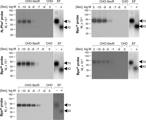
FIGURE 2.
Photoaffinity labeling of secretin receptor. Shown are representative autoradiographs of 10% SDS-polyacrylamide gels used to separate the products of affinity labeling membranes from CHO-SecR cells with each of the noted photolabile probes in the presence of increasing concentrations of competing unlabeled secretin (Sec; from 0 to 1 μm). As controls, labeling of the non-receptor-bearing CHO cell membranes by each probe in the absence of competitor is also shown. Each of the probes labeled the secretin receptor specifically and saturably with the labeling being competed by secretin in a concentration-dependent manner. The receptor bands labeled by each probe migrated at approximately Mr = 70,000 and shifted to approximately Mr = 42,000 after deglycosylation with endoglycosidase F (EF). No radioactive band was observed in the affinity-labeled non-receptor-bearing CHO cell membranes. Data are representative of at least three independent experiments.

