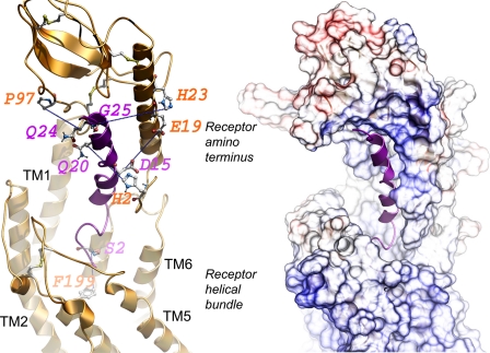
FIGURE 9.
Molecular model of ligand-bound secretin receptor. Shown is a lateral view of the best model of the secretin receptor-secretin peptide complex with the amino-terminal domain of the receptor above and the transmembrane helical bundle domain below. The secretin peptide ligand is illustrated in magenta with its carboxyl terminus within the peptide-binding cleft at the top and its amino terminus extending into the helical bundle at the bottom. The left panel highlights the five pairs of residues contributing to the experimental spatial approximation constraints (residues linked with blue dotted lines). The sites of incorporation of Bpa into the peptide ligand are identified in magenta, and the sites of covalent labeling of the receptor are identified in orange. The right panel illustrates the surfaces of the peptide-binding cleft within the receptor amino terminus and extending to the helical bundle.

