FIGURE 4.
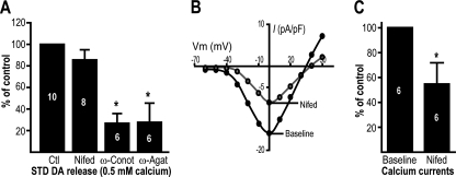
Somatodendritic DA release depends on N- and P/Q-type voltage-gated Ca2+ channels. A, 21-day-old DA neuron cultures were treated with 20 μm nifedipine (Nifed), 100 nm ω-conotoxin GVIA (ω-Conot), or 100 nm ω-agatoxin IVA (ω-Agat), and STD [3H]DA release was measured in the presence of 0.5 mm extracellular Ca2+. The graph represents the [3H]DA levels normalized against the control (Ctl, vehicle-treated) group. B, nifedipine (20 μm) efficiently blocks Ca2+ currents in DA neurons. Ca2+ currents, carried by barium, were measured in DA neurons cultured for 21 days. B and C, a representative Ca2+ current density (current size (pA)/membrane capacity (pF) ratio)-voltage plot is shown in B, whereas the measurement of peak Ca2+ current amplitude is summarized in C. The numbers inside the bars indicate the number of experiments performed in each group. *, p < 0.05 versus control. The error bars represent S.D.

