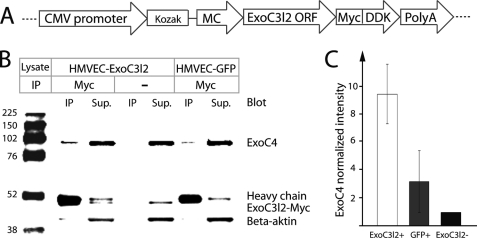
FIGURE 6.
EXOC3L2 physically interacts with EXOC4. A, the construct used to express Myc DDK-tagged human EXOC3L2 in endothelial cells (HMVEC). B, Western blot of co-immunoprecipitation (IP) of EXOC4 using the Myc-tagged EXOC3L2 as bait. The IP was also performed with no antibody or with HMVECs transfected with a GFP expressing plasmid (HMVEC-GFP) as negative controls. C, quantification of the data shown in B. The intensity of EXOC4 was normalized against intensity of β-actin in the supernatant and to the normalized intensity of the negative control. Error bars represent S.D. (n = 3).

