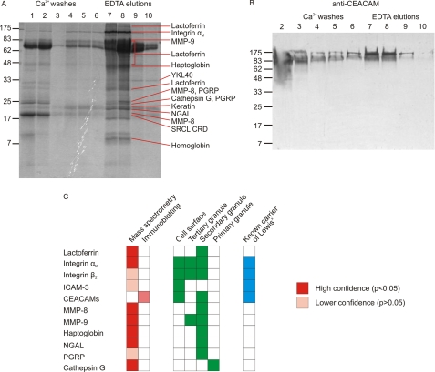
FIGURE 2.
Identification of SRCL ligands isolated from neutrophils. A, SDS-polyacrylamide gel of fractions from separation of granulocyte extracts on the SRCL affinity column. The gel was stained with Coomassie Blue, and slices were subjected to proteomic analysis. Major proteins identified in each band are indicated. Details of protein identification are provided in supplemental Tables S1 and S2. B, confirmation that some forms of CEACAM are ligands for SRCL. Blot of an SDS-polyacrylamide gel was probed with anti-CEACAM antibody, which was visualized with phosphatase-conjugated protein A. C, summary of ligands identified by proteomics and blotting. The distribution of glycoproteins in granulocytes is shown for comparison.

