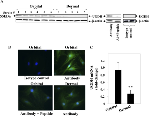
FIGURE 1.
UGDH protein and mRNA expression in orbital and dermal fibroblasts. A, immunoblot analysis of UGDH protein using an anti-UGDH polyclonal Ab. Cellular proteins from confluent orbital and dermal fibroblasts were solubilized, and equivalent amounts were loaded onto SDS-PAGE, transferred to a PVDF membrane, blocked, and probed with anti-UGDH Ab in a 1:1000 dilution. Pretreatment with UGDH peptide confirmed the Ab specificity by adsorbing any detectable immunoreactivity. A negative isotype control is shown on the far right. Membranes were then probed with β-actin as a loading control. B, UGDH was localized in both orbital and dermal fibroblasts by immunostaining with anti-UGDH Ab. Sub-confluent fibroblasts were grown on coverslips, fixed, blocked, and stained with anti-UGDH Ab without or with the UGDH peptide. Cells were then stained with an Oregon Green-conjugated goat anti-rabbit Ab, counter-stained, and mounted with Vectashield containing DAPI. Monolayers were visualized with an Axioskop40 microscope (Carl Zeiss, ×200). C, steady-state UGDH mRNA in orbital and dermal fibroblasts was quantified by real-time PCR. Confluent fibroblasts were shifted overnight to medium containing 1% FBS. RNA was extracted and reverse-transcribed. cDNA was subjected to quantitative real time-PCR. Each reaction was performed three times, and the values were normalized to their respective GAPDH signals and are expressed as the mean ± S.D. (**, p < 0.01).

